Animal Tissues Class 9

Table of Content

  • Animal Tissues
  • Epithelial Tissue
  • Connective Tissue
  • Muscular Tissue
  • Nervous Tissue
  • FAQs
  • Animal Tissues

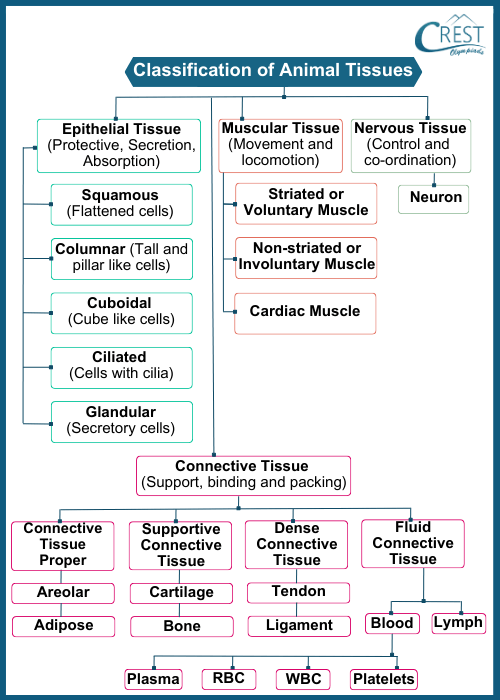
    Animal tissues are groups of specialised cells that work together to perform specific functions within an organism's body. These tissues are the building blocks of organs, and organs in turn work together to form complex systems that allow an organism to carry out various functions necessary for survival. Animal tissues vary in their structure, function, and origin, and they are categorised into four main types: epithelial, connective, muscle, and nervous tissues.

    Classification of Animal Tissues

    These tissues are further classified into various subtypes.

    Classification of Animal Tissues - Definition and Types etc

     Click to know more about Science Olympiad

    Epithelial Tissue

    Epithelial tissue plays a critical role in covering and protecting various parts of the body and its organs.

    Key Points

    1. Location and Function: Epithelial tissue covers the surfaces of organs and lines the cavities within the body. Its primary function is to create a barrier that separates different regions of the body or specific organs, protecting them from external factors, pathogens, and mechanical damage.
    2. Examples: Epithelial tissue is found in various locations, such as the skin, the lining of the mouth, the inner lining of blood vessels (endothelium), the digestive tract, and the respiratory tract.
    3. Cell Arrangement: Epithelial cells are tightly packed and closely adhere to each other, forming a continuous layer. This arrangement prevents substances from easily passing between the cells and helps maintain the integrity of the barrier.
    4. Function: Epithelial tissue serves as a protective shield and serves other functions as well, such as absorption, secretion, and sensory perception. It covers both external and internal surfaces of the body, acting as a selective barrier and allowing controlled interactions with the environment.

    Types of Epithelial Cells

    Epithelial cells, the building blocks of epithelial tissue, exhibit various types based on their shape, arrangement, and specialised functions.

    Based on Shapes

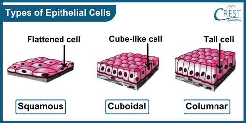
    1. Squamous Epithelium: These cells are flat and thin, resembling scales. They are found in tissues where substances need to move rapidly through barriers, such as the lining of blood vessels.
    2. Cuboidal Epithelium: Cube-shaped cells are found in glandular tissue and kidney tubules, where they contribute to secretion and absorption.
    3. Columnar Epithelium: These elongated, column-like cells are often specialised for absorption and have microvilli or cilia on their surfaces.
    4. Types of Epithelial Cells (Based on Shapes) - Definition, Function etc

    Based on their Arrangements

    1. Simple: Single layer of cells, suitable for surfaces where materials need to be absorbed or exchanged.
    2. Stratified: Multiple layers provide protection to underlying tissues and are found in areas subjected to wear and tear.
    3. Pseudostratified: While appearing layered, these cells are actually single-layered. Often found in respiratory passages, they have cilia for the movement of mucus.

    Based on their Types

    1. Simple Squamous Epithelium: Lines blood vessels and body cavities, regulating substance passage.
    2. Simple Cuboidal Epithelium: Found in glandular tissue and kidney tubules for secretion and absorption.
    3. Simple Columnar Epithelium: Lines the digestive tract, optimised for absorption, and often has microvilli.
    4. Stratified Squamous Epithelium: Offers protection on surfaces like the skin and oesophagus.
    5. Stratified Cuboidal Epithelium: Occurs in excretory ducts of glands.
    6. Stratified Columnar Epithelium: Lines areas like the conjunctiva, protective and mucus-secreting.
    7. Pseudostratified Columnar Epithelium: With cilia, lines respiratory passages, aiding in mucus movement.
     Read about Adaptation in Animals Based on their Eating Habits

    Based on Specialised Functions

    1. Transitional Epithelium: Stretches and contracts, allowing the urinary bladder to expand and contract.
    2. Glandular Epithelium: Found in glands, produces and secretes substances like hormones and enzymes.
    3. Olfactory Epithelium: In the nasal cavity, houses olfactory receptor cells with cilia to detect smells.

    Connective Tissue

    Connective tissue is one of the four primary types of animal tissues, along with epithelial, muscular, and nervous tissues. It serves a crucial role in the body by providing support, connecting different structures, and performing various other functions. Connective tissues are diverse in terms of structure, composition, and function, but they all share a common characteristic of having a non-living extracellular matrix that supports and surrounds the living cells within the tissue.

    Key Features of Connective Tissue

    1. Cells: Connective tissues contain a variety of cells, such as fibroblasts, adipocytes (fat cells), chondrocytes (cartilage cells), osteocytes (bone cells), and more. These cells are responsible for producing and maintaining the extracellular matrix.
    2. Extracellular Matrix: This matrix consists of a ground substance and fibres. The ground substance is composed of proteins and carbohydrates that create a gel-like or solid consistency. The fibres include collagen (provides strength), elastic (provides flexibility), and reticular (forms a network) fibres.
    3. Function: Connective tissues have multiple functions, including providing structural support to the body, connecting and anchoring organs, storing energy in the form of fat, transporting materials, and participating in immune responses and wound healing.

     Explore more about Micro-Organisms

    Types of Connective Tissues

    1. Loose Connective Tissue

    1. Description: Imagine a network of cells suspended within a flexible, jelly-like substance. It's like a soft cushion that surrounds and supports various body structures.
    2. Function: This tissue type is a bit like the body's "packing material." It provides support while allowing movement. It also acts as a storage site for fat cells and houses immune cells ready to defend against infections.
    3. Subtypes: Two main subtypes include areolar tissue (filling spaces between organs, allowing movement) and adipose tissue (storing energy as fat).

    2. Dense Connective Tissue

    1. Description: It is like a tough rope connecting things together.
    2. Function: This tissue provides firm connections between different body parts. Tendons attach muscles to bones, allowing movement, while ligaments connect bones to other bones, adding stability to joints.
    3. Subtypes: Regular dense connective tissue (parallel fibres, found in tendons and ligaments) and irregular dense connective tissue (random fibres, found in the skin's deeper layer).

    3. Supportive Connective Tissue

    1. Description: Think of a sturdy framework made up of specialised materials like cartilage and bone. It's like the scaffolding that holds things in place.
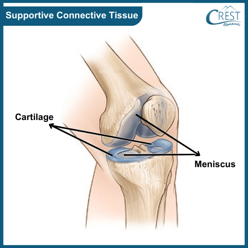
    2. Function: Cartilage is like a shock absorber, cushioning joints and maintaining structures like the nose. Bones, on the other hand, provide strength and rigidity to support the body and protect vital organs.
    3. Subtypes: Cartilage comes in various types such as hyaline (found in joints), fibrocartilage (in between spinal vertebrae), and elastic (in ears and epiglottis). Bones include compact (dense and strong) and spongy (spongy appearance, internal support) types.
    4. Diagram of Supportive Connective Tissue - Science Grade 9

    4. Fluid Connective Tissue

    1. Description: Picture cells suspended within a liquid medium, like fish swimming in water. Blood and lymph are the main players here.
    2. Function: Blood is like the body's courier service, transporting oxygen, nutrients, and waste products throughout. Lymph helps maintain fluid balance and supports the immune system.
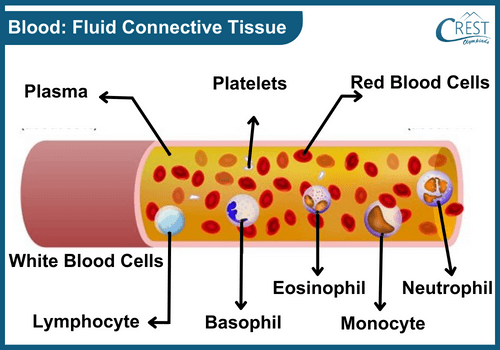
    3. Subtypes: Blood consists of red and white blood cells, and platelets, all suspended in plasma. Lymph is a colourless fluid containing immune cells, flowing through vessels and lymph nodes.
    4. Labelled Diagram of Blood: Fluid Connective Tissues - Definition, Subtypes and Functions etc

     Learn more about Reproduction in Animals

    Muscular Tissue

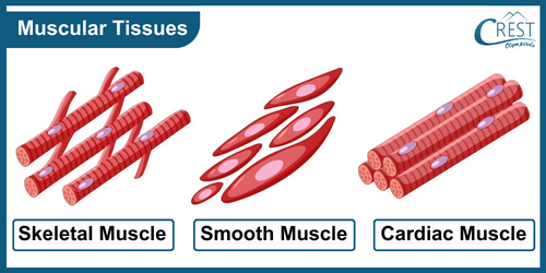
    Muscular tissue is a specialised type of tissue in our bodies responsible for generating movement, maintaining posture, and producing heat. It consists of elongated cells called muscle fibres or myocytes. These muscle fibres contain unique proteins that allow them to contract and relax, leading to movement.

    There are three main types of muscular tissue:

    Skeletal Muscle

    1. Also known as striated or voluntary muscle.
    2. It's attached to bones and is responsible for our conscious, voluntary movements like walking, jumping, and lifting.
    3. Skeletal muscle fibres are long, cylindrical, and multinucleated.
    4. The alternating pattern of light and dark bands, visible under a microscope, gives it a striated appearance.
    5. It's under conscious control, meaning we can choose when to contract or relax these muscles.

    Smooth Muscle

    1. Found in the walls of organs and structures like blood vessels, the digestive tract, and respiratory passages.
    2. It's involuntary, meaning we cannot consciously control its movements.
    3. Smooth muscle cells are spindle-shaped with a single nucleus.
    4. These muscles work to propel substances through various systems, like food through the digestive tract or blood through blood vessels.

    Cardiac Muscle

    1. Found exclusively in the heart.
    2. It's involuntary and responsible for the continuous pumping of the heart.
    3. Cardiac muscle cells are branched, interconnected, and possess a single nucleus.
    4. They contract rhythmically to pump blood throughout the body.
    5. Diagram of Types of Muscular Tissues - Science Grade 9

    Nervous Tissue

    Nervous tissue is a specialised type of tissue that plays a fundamental role in communication and coordination within the body. It's primarily composed of cells called neurons, which are capable of receiving, transmitting, and processing signals, also known as nerve impulses or electrical signals.

    Neurons

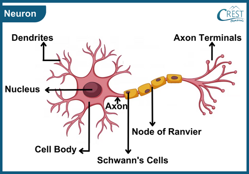
    1. Neurons are the functional units of nervous tissue.
    2. They possess a cell body containing the nucleus and other cellular components.
    3. Neurons have long, slender extensions called dendrites and axons.
    4. Dendrites receive incoming signals from other neurons or sensory receptors.
    5. The axon transmits these signals away from the cell body to other neurons or target cells.
    6. Labelled Daigram of Neuron - Nervous Tissue for Grade 9

    Structure of Neurons

    1. The cell body contains the nucleus and necessary cellular organelles.
    2. Dendrites are short, branching extensions that receive signals and convey them towards the cell body.
    3. The axon is a single, elongated projection responsible for transmitting signals away from the cell body.

    Function of Nervous Tissue

    1. Nervous tissue enables rapid communication between different parts of the body.
    2. It allows for sensory perception, motor control, coordination, and integration of various physiological processes.
    3. The brain, spinal cord, and peripheral nerves are composed of nervous tissue.

    Nervous System Components

    1. The central nervous system (CNS) includes the brain and spinal cord, where complex processing and integration of signals occur.
    2. The peripheral nervous system (PNS) comprises nerves that extend throughout the body to connect the CNS with muscles, glands, and sensory receptors.

    Frequently Asked Questions

    1. How does epithelial tissue protect the body from external damage?

    Epithelial tissue forms a protective barrier on the surface of the body and the lining of internal organs. It prevents the entry of pathogens, protects against mechanical injury, and reduces water loss from the body.

    2. Why is cardiac muscle unique among the types of muscle tissue?

    Cardiac muscle is unique because it is only found in the heart and has features of both smooth and skeletal muscles. It is involuntary, like smooth muscle, but has a striated appearance, like skeletal muscle. It is responsible for the continuous rhythmic contractions that pump blood throughout the body.

    3. Why is blood considered a connective tissue?

    Blood is considered a connective tissue because it consists of cells (red blood cells, white blood cells, and platelets) suspended in a liquid matrix (plasma). It connects different parts of the body by transporting oxygen, nutrients, and waste products.

    4. How does ciliated epithelial tissue function in the respiratory system?

    Ciliated epithelial tissue has hair-like structures called cilia on its surface. In the respiratory system, the cilia help move mucus and trapped particles out of the airways, keeping the lungs clean and preventing infections.

    5. What would happen if smooth muscle did not function properly in the digestive system?

    If smooth muscle did not function properly, the rhythmic contractions (peristalsis) required to move food through the digestive system would be disrupted. This could lead to problems with digestion and nutrient absorption.

    Quick Video Recap

    In this section, you will find interesting and well-explained topic-wise video summary of the topic, perfect for quick revision before your Olympiad exams.

    YouTube Video
    ×

    Share Your Feedback

    CREST Olympiads has launched this initiative to provide free reading and practice material. In order to make this content more useful, we solicit your feedback.

    Do share improvements at info@crestolympiads.com. Please mention the URL of the page and topic name with improvements needed. You may include screenshots, URLs of other sites, etc. which can help our Subject Experts to understand your suggestions easily.

    Other Science Related Topics for Class 9

    Other Subjects for Class 9

    70%